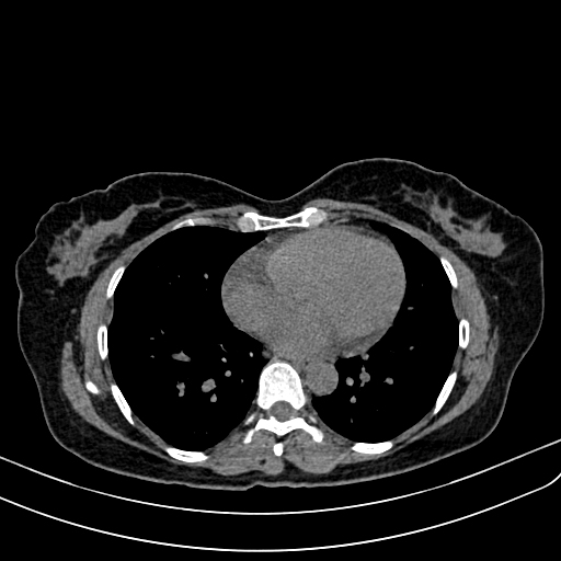
Incidental liver lesion on multiphase CT. Characterize the enhancement pattern before opening the answer.
Diagnosis
Cavernous hepatic hemangioma.
Key Observations
- Low attenuation on noncontrast imaging.
- Discontinuous peripheral nodular enhancement after contrast.
- Progressive centripetal fill-in on portal venous and delayed imaging.
- Enhancement persists rather than washing out.
Teaching conclusion: a classic hemangioma follows the blood pool over time; do not mistake progressive nodular fill-in for a rim-enhancing metastasis.