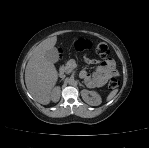
Focal liver lesion found after ultrasound. Use the noncontrast, arterial, portal venous, and delayed phases to characterize the enhancement.
Diagnosis
Hepatic hemangioma.
Key Observations
- Peripheral nodular enhancement after contrast.
- Progressive centripetal fill-in on later phases.
- The delayed phase supports benign blood-pool behavior rather than washout.
Teaching conclusion: progressive peripheral nodular fill-in is the classic dynamic pattern of a hemangioma.